ECR 2016 / C-1284
Eponyms in neurovascular anatomy
Congress:
ECR 2016
Poster Number:
C-1284
Type:
Educational Exhibit
Keywords:
Anatomy, Vascular, CNS, MR-Angiography, Catheter arteriography, CT-Angiography, Education, History, Embolism / Thrombosis, Obstruction / Occlusion
Authors:
A. Manzella1, L. Freitas2, H. Figueiredo2, B. Pernambuco2, G. Andrade3, B. Mota2; 1Recife, PE/BR, 2Recife/BR, 3RECIFE, PERNAMBUCO/BR
DOI:
10.1594/ecr2016/C-1284
. References: internet")
Fig. 1:
Franciscus Sylvius (1614-1672).
 of the middle cerebral artery. References: Hospital da Restauração")
Fig. 2:
Schematic drawing demonstrates the Sylvian branch (M2) of the middle cerebral...
 (green arrow). References: Hospital da Restauração")
Fig. 3:
Arteriography shows the same branch (M2) (green arrow).
. References: internet")
Fig. 4:
Johann O. L. Heubner (1843-1926).
 of the anterior cerebral artery (artery of Heubner). References: Hospital da Restauração")
Fig. 5:
Schematic drawing shows the recurrent branch (A2) of the anterior cerebral...
 demonstrates the artery of Heubner (yellow arrows). References: Hospital da Restauração")
Fig. 6:
Digital subtraction angiography (DSA) demonstrates the artery of Heubner...
. References: Internet")
Fig. 7:
Gerard Percheron (1930-2011).

Fig. 8:
Schematic drawing shows the artery of Percheron irrigating both sides of...
. Infarction of the artery of Percheron. References: Hospital da Restauração")
Fig. 9:
Diffusion weighted image at the level of the midbrain demonstrates hypersignal...
 at the level of the midbrain (infarction). References: Hospital da Restauração")
Fig. 10:
ADC map shows restricted diffusion (red arrow) at the level of the midbrain...
, in a patient with a vascular tumor in the tentorium region. References: Hospital da Restauração")
Fig. 11:
DSA demonstrates the Bernasconi-Cassinari artery (green arrow), in a patient...
. References: Hospital da Restauração")
Fig. 12:
DSA shows a healthy individual with the tentorial artery (yellow arrow).
. References: Internet")
Fig. 13:
Schematic drawing shows the Bernasconi-Cassinari branch (black arrow).
. References: Internet")
Fig. 14:
Vidius Vidius (1509-1569).

Fig. 15:
Schematic drawing of the the Vidian artery in the Vidian canal.
 shows the pterygoid canal (green arrow). References: Hospital da Restauração")
Fig. 16:
Axial CT (bone window) shows the pterygoid canal (green arrow).
. References: Hospital da Restauração")
Fig. 17:
DSA demonstrates the Vidian artery (yellow arrow).
. References: Internet")
Fig. 18:
Thomas Willis (1621-1675).

Fig. 19:
Schematic drawing shows the arteries that form the polygon of Willis. MCA:...
. 3D reconstruction shows the arteries of the circle of Willis. References: Hospital da Restauração")
Fig. 20:
MR angiography (MRA). 3D reconstruction shows the arteries of the circle of...
. References: Internet")
Fig. 21:
Albert W. Adamkiewicz (1850-1921).

Fig. 22:
Illustration demonstrates two different levels of the origin of Adamkiewicz...
. References: Hospital da Restauração")
Fig. 23:
DSA demonstrates the artery of Adamkiewicz (green arrow).
. References: Internet")
Fig. 24:
Friedrich C. Rosenthal (1780-1829).
. References: Hospital da Restauração")
Fig. 25:
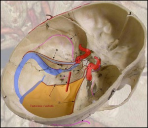
Schematic representation of the venous circulation of the brain. The basal vein...
 shows the venous circulation of the brain. Basal vein of Rosenthal (arrow). References: Hospital da Restauração")
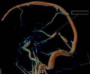
Fig. 26:
MRA (3D reconstruction) shows the venous circulation of the brain. Basal vein...
 demonstrates the basal vein of Rosenthal. References: Hospital da Restauração")
Fig. 27:
DSA (venous phase) demonstrates the basal vein of Rosenthal.
. References: Internet")
Fig. 28:
Galen of Pergamum (129-210 B.C).
 demonstrates the venous circulation of the brain. Vein of Galen (arrow). References: Hospital da Restauração")
Fig. 29:
MRA (3D) demonstrates the venous circulation of the brain. Vein of Galen...
 shows the great cerebral vein (green arrow). References: Hospital da Restauração")
Fig. 30:
DSA (venous phase) shows the great cerebral vein (green arrow).

Fig. 31:
Schematic representation of the Charles Labbé`s doctoral paper in 1879...
. Vein of Labbé (white arrow). References: Hospital da Restauração")
Fig. 32:
DSA (venous phase). Vein of Labbé (white arrow).
. Vein of Labbé (arrow). References: Hospital da Restauração")
Fig. 33:
MRA (3D). Vein of Labbé (arrow).
. References: Internet")
Fig. 34:
Jean B. P. Trolard (1842-1910).
. Vein of Trolard (arrow). References: Hospital da Restauração")
Fig. 35:
MRA (3D). Vein of Trolard (arrow).
. References: Internet")
Fig. 36:
Herophilus (340-280 B.C).
. Torcula herophili (gree arrow). References: Hospital da Restauração")
Fig. 37:
DSA (venous phase). Torcula herophili (gree arrow).
. Confluence of venous sinus (Torcula herophili). References: Hospital da Restauração")
Fig. 38:
MRA (MIP). Confluence of venous sinus (Torcula herophili).
. References: Internet")
Fig. 39:
Walter E. Dandy (1886-1946).
 shows the vein of Dandy (green arrow). References: Hospital da Restauração")
Fig. 40:
DSA (venous phase) shows the vein of Dandy (green arrow).

Fig. 41:
HOSPITAL DA RESTAURAÇÃO
RECIFE - PERNAMBUCO - BRAZIL